

Mammogram
Description
 Mammography is a specific type of imaging that uses a low-dose x-ray system and
high-contrast, high-resolution film for examination of the breasts. A mammography
unit is a rectangular box that houses the tube in which x-rays are produced. The
unit is dedicated equipment, because it is used exclusively for x-ray exam of the
breast with special accessories that allow only the breast to be exposed to the
x-rays.
Mammography is a specific type of imaging that uses a low-dose x-ray system and
high-contrast, high-resolution film for examination of the breasts. A mammography
unit is a rectangular box that houses the tube in which x-rays are produced. The
unit is dedicated equipment, because it is used exclusively for x-ray exam of the
breast with special accessories that allow only the breast to be exposed to the
x-rays.
Attached to the unit is a device that holds and compresses the breast and positions it so images can be obtained at different angles.
Mammography plays a central part in screening for breast cancers because it can show changes in the breast up to two years before a patient or physician can feel them. Mammography is used to diagnose breast diseases in women.
Initial mammographic images themselves are not always enough to determine the existence of a benign or malignant disease with certainty. If a finding or spot seems suspicious, the radiologist may recommend further diagnostic studies.
Procedure
No preparation is required.
The breast is exposed to a small dose of radiation to produce an image of internal breast tissue. The image of the breast is produced as a result of some of the x-rays being absorbed (attenuation) while others pass through the breast to expose the film. The exposed film is placed in a developing machine to produce images.
Precautions
Patients should not wear deodorant, talcum powder, or lotion under their arms on the day of the exam. These can appear on the x-ray film as calcium spots.
All jewelry and clothing above the waist should be removed.
Location
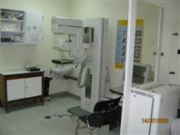
There is 1 room located at the Radiology Department - SMC New Building - Ground Floor.
Tel. No.: 17284007
Ext. No. : 4007
Equipment
SMC has one digital mammography unit with biopsy digital (Seno Essential) manufacture by GE. This unit has workstation with compute-aided detection (CAD).
CAD it is software analyzes the digital representation of the mammogram image and marks suspicious areas on the screen image for the radiologist to review in association with their own reading of the original images. Preliminary studies suggested that the device helped in detection of cancer. Many organizations such as insurance companies are reimbursed for the application of CAD. The software nowadays has been spread into practice, and it is estimated that about 30 percent of mammograms are now interpreted using one of these devices.
N.B. The old unit was call Mammomat 3000- Optima and it was manufactured by Siemens.
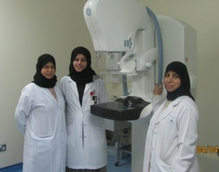
Available Staff
Three Consultant Radiologists: Dr. Herminia AlSaffar is in charge of the breast imaging service supported by Dr. Neelam Malik and Dr. Jamila Al.Dosary.
Dr.Marwa Chief Resident (Breast Imaging followship).
One senior Radiographer (per shift): Mrs. Badriya Al.Najjar / Mrs. Zahra Majeed/ Mrs.Noora Farhan
Or one radiographer Mrs. Najat Ali and Mrs. Danya
Timing
Sunday to Thursday from 7:00 AM to 2:00 PM