

Magnetic Resonance Imaging (MRI)
Description
Magnetic Resonance Imaging (MRI) has evolved into one of the most powerful non-invasive
techniques in diagnostic imaging. The MRI examination represents a new way of looking
at disease processes without the use of radiation.
Instead, magnetic resonance is used to provide images, which provide excellent anatomical
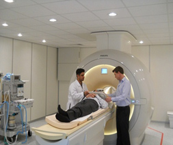
detail and reflect changes in tissue chemistry. During the exam, the patient lies
on a table in a strong magnetic field while a computer collects the information
used to create images.
MRI employs the magnetism of the hydrogen nucleus, the proton, and its interaction with external magnetic and electromagnetic fields. It produces cross-sectional pictures like computed tomography, without the use of any harmful ionizing radiation. MRI images are due to differential proton densities of the tissues in a layer of the body under study.
MRI is a diagnostic imaging technology that uses a strong magnet and radiofrequency waves to produce pictures or "images" of internal organs and structures. Because MRI allows us to see inside the body from any angle with great clarity, it gives us a wealth of information more quickly and in many cases, more economically than past tests and exploratory surgeries. The examination may take from 15 to 30 minutes.
MRI is the preferred method of imaging the brain to detect the presence of ischemia,
inflammation, demyelination, tumors and congenital anomalies. Due to the excellent
tissue contrast of MR, MRI provides better tissue contrast compared to CT scan.
Other applications include: spinal, musculoskeletal, abdominal, pelvic and breast
imaging.
Over 6800 MRI scans are performed at SMC per year.
Pre-procedure Instructions/Preparation
MR technologists must know if the patient has a pacemaker, surgical clips, a prosthesis, metal implants or any other metal objects in the body. Some implants (e.g., a pacemaker) may be affected by an MR examination. The MRI staff will then determine whether or not the patient should proceed with the MR examination.
Any metal materials that might be affected or attracted by the powerful magnet used for MR imaging should be left at home or given to the MR staff for safekeeping. This list includes coins, keys, bobby pins, credit cards, pocketknives, Hearing aids, dentures, watches, hair wigs or hairpieces, artificial eyes, glasses, jewellery, earrings, etc. Patients should also be certain that they are reasonably clean from any eye make-up or as a result of working around metal finishing or grinding equipment. Contrast material may be given by intravenous injection, but generally there are no dietary restrictions and all medications may be taken without interference.
Preparation
It is advisable to exclude women during the first three months of pregnancy.
Empty bladder before examination.
Procedure
For MR imaging, the patient is placed in a strong magnetic field, which causes the nuclear magnetism of protons of hydrogen atoms to become aligned. A hydrogen atom contains a single proton in its nucleus and is the most abundant element in the body. As it has a strong magnetic component, it is the ideal choice for live imaging.
Then a radio frequency pulse at resonant frequency is transmitted to the patient.
A radio signal generated by hydrogen nuclei present in the body cells under the
influence of a strong magnetic field is detected, which is ultimately processed
in the form of a tomographic image by high-tech inbuilt computers. In most MRI instruments,
niobium-titanium superconducting magnets are used.
The procedure will take approximately 45 minutes to complete. Outpatients are to
arrive in out patient registration 45 minutes prior to the exam for registration.
Children may require sedation for the procedure. If this is necessary, special arrangements
will be made by the referring physician before the visit.
Precautions
Patients with cardiac pacemakers, artificial heart valves, aneurysm clips in the brain, surgicals, implants, prosthetic devices cannot be scanned unless the implants are MRI-compatible. Scanning is generally avoided in pregnancy (especially during the first trimester).
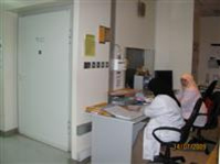
Location and Contact
There are two MRI units, both of them they are located opposite to SMC pharmacy in SMC old Building - Ground Floor.
MRI No.1 which is 3 Tesla unit.
Tel. 17285871 Ext. 5871
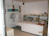
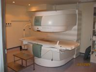
The second one is an open MRI (0.35 Tesla). Both of them they are located on the main corridor near the main Pharmacy.
Tel. 17285054 Ext: 5054
MRI Appointment office 17285873 Ext. No: 5873
Equipment
There are two MRI scanners in SMC.
One AchievaTX MRI unit it is the first 3T MRI unit to be installed in GCC region which is rated as the best in the industry standards. Achieva TX is with Multi-transmit, the state of the art technology which delivers enhanced clinical performance in a wide range of applications. This unique system enables excellent body imaging as well as ultra fast scanning and improves lesion visualization significantly. Besides multi-transmit the system embodies all the pioneering technology that has made Philips Achieva TX system the benchmark in high field MRI with the largest installed base of compact 3T systems in the world.
The 2nd unit is open Magnet 0.35T it is manufactured by Siemens.
MRI equipment is very expensive unit. Hence the cost of every scan is very high. The installation cost of MRI equipment is very high, as it requires an A/C chamber which will not cause any hindrance in the magnetism of the instrument. It is difficult and sometimes impossible to perform an MRI scan on a person with some metallic implants like a dental implant, metal rods, pacemaker or artificial heart valves.
In spite of this, MRI can be considered as one of the most revealing diagnostic aids available today.
N.B. the old MRI was 1 Tesla and it is (Magnetom - Impact - 1.0Tesla it was made by Siemens).
Available Staff
-
Consultant Radiologists:
- Dr. Fatima A.Rahman (MSK MRI).
- Dr. Suzanne Abbas (Neuro MRI).
- Dr. Hakima Al.Hashimi (Paediatric MRI).
- Dr. Sharif Al.Arrayedh (Body MRI).
- Dr. Husain Nasser (Paediatric MRI).
- Dr. Jameela Al Dossary (Spine MRI).
- Dr. Mona (GYN MRI).
- Dr. Marwa (Breast, GGM MRI).
- Dr. Herminia (Breast MRI).
- The in-charge of: MRI is Mrs. Zahra Makki (Senior Radiographer)
- Radiologic Technologists Mr. Abdulla Hasan
- Four Senior Radiographers : Mrs. Ebtisam Radi, Mr. Rashid Al.Aradi, Mrs. Amira Hasan and Mrs. Amal Rashed
- Please refer to the technologist and radiologist on call list for out-of-hour emergencies.
- One staff nurse: Miss Jehan
Timing
Sunday to Thursday from 7:00 AM to 2:00 PM
+24 hours Radiologists & Radiographers on-call