
Fluoroscopy and Special Procedures
Description
 The most common fluoroscopy procedures are (upper GI series)
Ba. Meal, Barium swallow, Small Bowel Meal (SBM), and Barium Enema. Fluoroscopy
can be thought of as an X-Ray movie. During this examination, the radiologist watches
Barium in different parts of the digestive tract and takes a series of X-Ray images
during the exam.
The most common fluoroscopy procedures are (upper GI series)
Ba. Meal, Barium swallow, Small Bowel Meal (SBM), and Barium Enema. Fluoroscopy
can be thought of as an X-Ray movie. During this examination, the radiologist watches
Barium in different parts of the digestive tract and takes a series of X-Ray images
during the exam.
For a Barium Swallow, Ba. Meal and SBM, the patient drinks a Barium liquid which
allows the radiologist to see the inside of the esophagus, stomach and intestine.
Frequently, the patient will also have to take gas granules, which distend the esophagus
and stomach. For a Barium Enema, a tube is inserted into the rectum through which
Barium liquid is administered to fill the colon. This is frequently followed by
the administration of air. This exam allows the radiologist to see the inside of
the colon. A Barium swallow, Ba. Meal and Barium Enema require around 45 minutes
to an hour. A SBM can take one and half hour to 3 hours depending upon how fast
the Barium moves through the small intestine.
Procedure
Most special procedures require patient preparation before the examination. Hence they must be given an appointment prior to the examination.
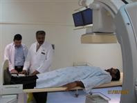
In Most Fluoroscopy or special procedure examinations, the patient lies on an X-Ray table during his/her examination.
Barium Meal (Double contrast): now used by many radiologists routinely
as it is particularly useful to demonstrate the mucosal pattern of the stomach and
duodenal cap more clearly and thus detect small lesions at an early stage. Not recommended
for children or very ill adults. It may be carried out with or without the injection
of a muscle relaxant to produce gastric atony.
Technique: in the erect position under screen control a small mouthful of barium
is given followed by a gas producing agent with a further small amount of barium.
The patient is then placed supine and is rotated through 360° to coat the gastric
mucosa. Films are taken in various positions.
IVU (intravenous urography)
In this, a dye is injected intravenously and x-ray images of the kidneys, ureters
and bladder are obtained. The dye is radio-opaque and seen well with x-rays. Overnight
fasting and good preparation of the colon with Dulcolax tablets are required to
be taken by the patient two days before the appointment.
MCU (micturating cystourethrography)
Dye is introduced into the urinary bladder through a catheter and the patient is
asked to micturate/urinate. X-ray images are obtained during the act of micturition
to assess the function and structure of the urinary bladder and urethra.
RGU (retrograde urethrography)
Dye is injected through the urethra from the penis and x-ray images are taken. This
helps in assessing the urethra and the bladder base.
Fistulogram and sinusogram
In these studies, using a small catheter, iodinated dye is injected into the cutaneous
sinus or fistula and x-rays are taken, which help in identifying the tract of the
sinus or fistula.
Sialography
In this, the parotid or submandibular duct is cannulated from the mouth and x-ray
images of the parotid or submandibular ducts and gland are obtained.
HSG (Hysterosalpingography)
The cervix is cannulated and iodinated dye is injected into the cervical and uterine
lumen. The Fallopian tubes are then well seen. This procedure is used to study the
patency of the passage as well as other structural abnormalities.
Apart from the routine studies mentioned above, other special procedures can be carried out in the department such as:
- Myelogram (only if strongly indicated).
- Postoperative (T-Tube) cholangiography.
- Percutaneous transhepatic cholangiography (PTC).
- Dacrocystogram.
- Venogram.
- Fluoroscopy for any part of body (e.g. for diaphragmatic movement).
- Some of those examinations require patient preparation and others do not.
Precautions
Scanning is NOT allowed for pregnant women.
Patients undergoing IVU's should be checked for allergies and renal functions.
Location
There are 3 rooms (Rooms 3, 4 and 5) located at the Radiology Department - SMC Old
Building - Ground Floor.
Fluoroscopy examinations are done in rooms 3 and 4 while IVU's are performed in
room 5.
Tel. No.: 17285018 / 17285538
Ext. No. : 5018 / 5538
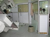
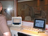
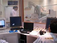
Equipment
We have two digital Fluoroscopy systems in SMC radiology department, X-Ray rooms,
No. 3 and 4. Room No. 4 is equipped with digital R/F (Diagnost 96) system
manufactured by Philips.
Around 70 % of the Fluoroscopy procedures are done on this system because the radiologist
can stand behind a lead screen or in the control area which means less radiation
for radiology staff compared to other systems. This unit is fully remote controlled
and is fully digital with spot film device.
Room No.3 is equipped with MultiDiagnost Eleva with flat detector,it is manufactured by Philips.
The MultiDiagnost Eleva is multi-purpose system suitable for both digital fluoroscopy and imaging. It is, therefore, ideal for a wide range of routine and special examinations requiring special patient positioning and compound X-ray beam projections. It is consists of a floor mounted X-ray stand with tilting tabletop. The stand's main support beam carries both the tabletop support the C-arc. The C-arc allows the flat detectors and X-ray detector and X-ray tube to move around the patient.
In this system the radiologist and the radiographer have to be behind the control panel and incase the staff need to be stand inside control area he/she has to wear lead protection aprons, lead thyroid shields and lead glasses if wanted.
The units are connected with dry laser printers either Agfa or Kodak for printing patients images.
These systems have a broad range of image-optimizing components. They provide for high quality images at minimum dose, allowing us to make diagnoses with a high degree of confidence.
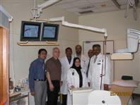
Available Staff
Each of those rooms is operated by one radiographer.
Almost all of the special procedures are performed by a consultant or a resident
radiologist with the aid of a radiographer or technologist.
Timing
Sundays to Thursdays from 7:00 AM to 2:00 PM
+24 hours Radiologists & Radiographers on-call