
Endoscopic Retrograde Cholangiography and Pancreatography (ERCP)
Description
ERCPs were preformed in the Radiology Department 8 years ago under the Fluoroscopy
system. The procedure has since been shifted to the Endoscopic Department on the
3rd level at SMC.
ERCP is a valuable examination for the diagnosis of many diseases of the pancreas,
bile ducts, and gallbladder. ERCP allows the doctor to perform necessary treatments
such as dilating the CBO, removing gallstones lodged in the bile duct, inserting
a stent (drain) in the duct or taking a biopsy specimen (tiny bit of tissue).
Procedure
A flexible fiberoptic/video tube (duodenoscope) is passed through the
mouth, esophagus (food tube) and stomach into the duodenum (second part of the small
intestine). The ampulla (opening where the bile and pancreatic ducts empty into
the duodenum) is then identified.
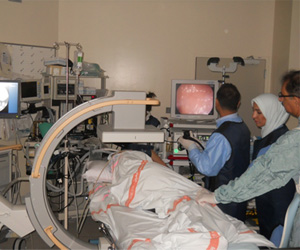
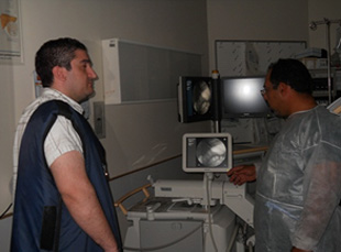
A small plastic tube (canula) is passed through the duodenoscope into the
ampulla. X-ray dye is injected through the canula into the ducts. With the help
of X-rays fluoroscopy system images are taken for the study of the ducts. Any necessary
treatments can be performed at this time.
Patients should remove dentures and eyeglasses prior to the start of the procedure.
They are asked to sign a consent form authorizing the doctor to perform the procedure.
Patient should tell the doctor or the GI nurse if he/she is allergic to any medicines
or X-Ray Contrast media.
A needle for intravenous (IV) medicines and fluids will be placed in the patient's arm vein. Medicine will be injected through the IV needle that will make the patient sleepy and relaxed. The doctor may spray the patient's throat or ask him/her to gargle with a numbing medicine.
The patient will lie on an x-ray table on his/her left side and a small plastic mouthpiece will be placed between his/her teeth. The patient will be able to breathe normally. The doctor will help him/her to swallow the lubricated flexible duodenoscope tube. When the tube is present in the duodenum, the patient will be helped to turn onto his/her abdomen with his/her head turned to the right.
During the procedure, the patient may feel some abdominal fullness or bloating due to the air which the doctor puts into the duodenum. As the x-ray dye is injected into the ducts, the patient may feel some mild discomfort. These feelings should be completely tolerable and not painful. ERCP takes 30 minutes to 2 hours. After the procedure, the patient will need to stay at the hospital for 1 to 2 hours until the sedative wears off.
After the duodenoscope is removed, the patient may be asked to move into various positions so that more x-rays can be taken.
Many people do not recall any of the procedure because of the effect of the medicine. After the procedure the patient will probably feel drowsy and may sleep for a short time.
Approximately 89 routine ERCP procedures are performed annually at SMC using the mobile C. arm fluoroscopy unit in the endoscopic department.
Precautions
Scanning is NOT allowed for pregnant women.
Location and Contact
The Endoscopy suite at SMC is located on the 3rd floor - New Building. The suite currently has 3 procedure rooms. Only one of these rooms is allocated for ERCP as it is shielded.
Dir. Tel. No.: 17284675
Ext. No.: 4675 -4677
 Equipment
Available Staff
We usually send one Radiographer to cover the examinations.
One Consultant Physician & Gastroentrologist:
Dr. Jehad Radhi Al-Qamish, MBCh. MRCP (UK), FRACP
Endoscopy nurses:
Sister Majeda & Sister Kawakab
Timing
Mondays only from 7:00 AM to 2:00 PM
At present there are no facilities available to perform the procedure other than
during daily working hours due to lack of manpower.
In case for emergency we will arrange one radiographer to work with ERCP team.