

Angiography (Vascular and Interventional)
Description
Angiography is the x-ray study of the blood vessels. An angiogram uses a radiopaque
substance, or dye, to make the blood vessels visible under x ray. Arteriography
is a type of angiography that involves the study of the arteries. Angiography is
used to detect abnormalities or blockages in the blood vessels (called occlusions)
throughout the circulatory system and in some organs.
The procedure is commonly used to identify atherosclerosis; to diagnose ischemic
heart disease; to evaluate tumors; to detect an aneurysm (an abnormal bulge of an
artery that can rupture leading to hemorrhage), dissections, blood clot and arteriovenous
malformations (abnormal tangle of arteries and veins).
Procedure
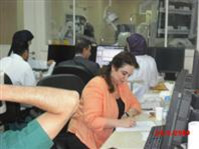
Angiography is usually performed at a hospital by a trained radiologist and assisting technician or nurse. It takes place in an x-ray or fluoroscopy suite, and for most types of angiograms, the patient's vital signs will be monitored throughout the procedure. Angiography requires the injection of a contrast dye that makes the blood vessels visible to x ray. The puncture is usually made in the groin area, armpit, inside elbow, or neck. The site is cleaned with an antiseptic agent and injected with a local anaesthetic. A fluoroscopic screen that displays a view of the patient's vascular system is used to see the correct location. The patient is warned that when the injection starts, he/she should remain very still. The injection causes some mild to moderate discomfort. Possible side effects or reactions include headache, dizziness, irregular heartbeat, nausea, warmth, burning sensation, and chest pain, but they usually last only momentarily. To view the area of study from different angles or perspectives, different injection will be made and the patient or the camera maybe needed to move.
Precautions
Because X rays carry risks of ionizing radiation exposure to the fetus, pregnant women are also advised to avoid this procedure.
Patients with kidney disease or injury may suffer further kidney damage from the
contrast mediums used for angiography. Patients who have blood clotting problems,
have a known allergy to contrast mediums, or are allergic to iodine, a component
of some contrast mediums, may also not be suitable candidates for an angiography
procedure.
Location and Contact
There are two units in SMC one in room No. 6 which is located at the Radiology Department
- SMC Old Building - Ground Floor
X-Ray room 6 Ext. No. : 5694 Dir. 17285694
Ward 11 Angiogram room Ext. No.: 7807 Dir. 17287807 Ext.7814 Dir.17287814
This unit is located in ward 11 on ground floor old SMC building
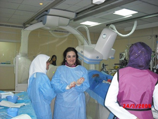
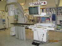
Equipment
One bi plane unit and it is GE manufactured. This unit is located in ward 11 on ground floor old SMC building.
There are plans to install a second unit in room 6 in the main radiology department in the near futur.
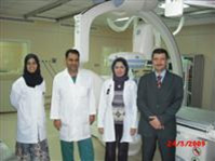
Available Staff
Consultant Radiologists:
Dr. Shareef AlArrayed (majority of vascular and interventional),
Dr. Suzanne Abbas (neuro-vascular)
One Resident Radiologist: (see the radiologist list)
Head of Vascular & Interventional Unit
Mrs. Karima Hassan
Two Radiographers (per shift):
A.Ali Mahfoodh / Alawi S.Saeed /Muntaha Hassan /Abbas Mahdi
3 staff Nurses : Ahlam Jumma (BSc), and Fatima Ali (BSc).
Timing
Sunday to Thursday from 7:00 AM to 2:00 PM
24 hours Radiologist & Radiographer on-call